Summary / Overview
No key-points marked yet. Add lines like *Important point* in this section.
Etiology
No key-points marked yet. Add lines like *Important point* in this section.
Pathogenesis
No key-points marked yet. Add lines like *Important point* in this section.
Symptoms
No key-points marked yet. Add lines like *Important point* in this section.
Signs
No key-points marked yet. Add lines like *Important point* in this section.
Clinical Features
No key-points marked yet. Add lines like *Important point* in this section.
Investigations
No key-points marked yet. Add lines like *Important point* in this section.
Differential Diagnosis
No key-points marked yet. Add lines like *Important point* in this section.
Complications
No key-points marked yet. Add lines like *Important point* in this section.
Treatment
No key-points marked yet. Add lines like *Important point* in this section.
Prevention
No key-points marked yet. Add lines like *Important point* in this section.
Serotypes / Subtypes
No key-points marked yet. Add lines like *Important point* in this section.
Pathology
No key-points marked yet. Add lines like *Important point* in this section.
Radiology / Imaging
No key-points marked yet. Add lines like *Important point* in this section.
Notes / Teaching points
No key-points marked yet. Add lines like *Important point* in this section.
Upper respiratory illness predominates in most patients
Children commonly present with croup (laryngotracheobronchitis)
Barking cough + hoarseness are characteristic
Stridor indicates upper airway obstruction
Nasal congestion + rhinorrhoea frequent early features
Fever usually mild–moderate; high fever uncommon
Lower respiratory involvement → bronchiolitis or pneumonia
Wheezing and dyspnea in infants and elderly
Symptoms more severe in immunocompromised patients
Seasonal outbreaks — especially in pediatric populations
Recurrent infections occur; immunity is incomplete
Acute laryngotracheobronchitis (croup) — most common complication in children
Airway obstruction due to subglottic edema
Bronchiolitis in infants
Secondary bacterial infection — otitis media, sinusitis, pneumonia
Dehydration due to poor feeding and fever
Hypoxia in severe respiratory involvement
Respiratory failure (rare; severe pediatric or immunocompromised cases)
Recurrent wheezing in susceptible children after infection
Influenza (A/B/C) — higher fever, systemic toxicity more prominent
Respiratory Syncytial Virus (RSV) — bronchiolitis predominant in infants
Adenovirus infection — conjunctivitis + pharyngitis common
COVID-19 — systemic symptoms, anosmia, pneumonia patterns
Bacterial tracheitis — high fever, toxic child, purulent secretions
Epiglottitis — sudden onset, drooling, severe airway obstruction
Diphtheria — membranous pharyngitis, neck swelling
Allergic rhinitis — no fever, eosinophilic inflammation
Foreign body aspiration — sudden onset stridor without prodrome
Pertussis — paroxysmal cough with inspiratory whoop
Human parainfluenza viruses (HPIV) are the causative agents
• Family: Paramyxoviridae
• Enveloped, single-stranded negative-sense RNA viruses
• Spread mainly via respiratory droplets and direct contact
Four major human types
• HPIV-1 → most common cause of croup outbreaks
• HPIV-2 → croup and URTI
• HPIV-3 → bronchiolitis and pneumonia (infants)
• HPIV-4 → milder respiratory illness, less common
Primary mode of transmission
• Respiratory droplets (cough/sneeze)
• Contaminated surfaces → hand-to-nose/eye transmission
• Close contact settings (households, daycare, hospital)
High-risk groups
• Infants and young children
• Elderly
• Immunocompromised patients
• Chronic lung disease patients
Seasonal pattern
• HPIV-1 → autumn outbreaks
• HPIV-2 → late autumn
• HPIV-3 → spring and early summer
• HPIV-4 → variable, less defined seasonality
Not a bacterial disease
• Secondary bacterial infection may occur but virus is primary cause.
Diagnosis is primarily clinical in mild upper respiratory infections
RT-PCR from nasopharyngeal swab — most sensitive and specific test
Viral antigen detection (DFA/rapid tests) — supportive but less sensitive
Multiplex respiratory viral panel — identifies parainfluenza with other viruses
CBC usually normal or mild lymphocytosis
CRP/ESR mildly elevated in inflammatory cases
Chest X-ray if LRTI (lowerRespiratoryTractInfection) suspected — peribronchial thickening, hyperinflation
Neck X-ray (croup) — steeple sign due to subglottic narrowing
Pulse oximetry — assess hypoxia in moderate–severe disease
ABG in severe respiratory distress
Parainfluenza is the most common viral cause of croup in children
Croup age group: 6 months – 3 years (peak incidence)
Barking cough + inspiratory stridor strongly suggests laryngotracheitis
Severity depends on degree of subglottic edema rather than viral load
Symptoms often worse at night due to circadian airway reactivity
Cool mist and calm environment reduce airway spasm in children
Steroids are the mainstay of treatment in moderate–severe croup
Nebulized adrenaline provides rapid but temporary relief in airway obstruction
Antibiotics are NOT indicated unless secondary bacterial infection suspected
Recurrent croup may suggest airway hyperreactivity or structural anomaly
Adults usually develop mild URTI; severe airway disease uncommon
Immunity is incomplete — reinfections occur throughout life
Virus enters through upper respiratory mucosa via inhaled droplets
Initial replication occurs in nasal and nasopharyngeal epithelial cells
Spread to larynx, trachea, and bronchi leads to inflammatory airway edema
Croup results from subglottic mucosal edema and narrowing of pediatric airway
Bronchiolitis develops due to infection of small airway epithelium
Pneumonia occurs when infection extends to lower respiratory tract and alveoli
After mucosal entry, virus attaches to epithelial cells via hemagglutinin-neuraminidase proteins.
Local viral replication triggers epithelial cell injury and mucociliary dysfunction.
Host immune response activates cytokines → IL-6, interferons, TNF-α
Inflammatory cell infiltration leads to airway obstruction and wheeze
Severe disease occurs in infants due to narrow airway caliber
Immunocompromised hosts show prolonged viral replication and severe LRTI
Secondary bacterial infection may follow epithelial damage in severe cases.
Virus infects respiratory epithelial cells of upper and lower airway
Attachment via hemagglutinin-neuraminidase (HN) protein to host cells
Fusion protein (F) mediates viral entry and syncytium formation
Epithelial cell necrosis and desquamation occur in infected airway
Submucosal edema develops due to inflammatory response
Perivascular and peribronchial lymphocytic infiltration seen
In croup — marked subglottic mucosal edema causes airway narrowing
In bronchiolitis — small airway obstruction due to inflammation + mucus plugging
Goblet cell hypersecretion increases mucus production
Vascular congestion contributes to mucosal thickening
Severe cases may show diffuse lower respiratory tract involvement
No chronic structural lung damage in uncomplicated cases
No licensed vaccine currently available for routine prevention
Droplet transmission is the main mode → respiratory hygiene is key
Frequent hand washing reduces viral spread
Avoid close contact with symptomatic individuals
Isolation of infected children in hospital/ward settings
Mask use during outbreaks reduces transmission
Surface disinfection in daycare and pediatric wards
Avoid sharing utensils, towels, toys during illness
High-risk groups require protection:
Infants
Elderly
Immunocompromised patients
Breastfeeding provides partial passive immune protection in infants
Community awareness during seasonal outbreaks helps early containment
Neck X-ray (AP view) in croup shows “steeple sign” due to subglottic narrowing
Lateral neck film may show airway narrowing without epiglottic swelling
Chest X-ray usually normal in mild URTI cases
Perihilar infiltrates may be seen in lower respiratory involvement
Hyperinflation seen in bronchiolitis due to air trapping
Patchy atelectasis may occur from mucus plugging
No focal lobar consolidation unless secondary bacterial infection
Ultrasound rarely used — may show airway narrowing in pediatric settings
CT chest not routinely required — used only in severe/complicated cases
References
PDF
cureus.com
2026-02-22 23:17:10
Human Parainfluenza Virus (HPIV) belongs to Paramyxoviridae family
Enveloped, single-stranded negative-sense RNA virus
HPIV-1 — most common cause of croup (autumn outbreaks)
HPIV-2 — also associated with croup but less frequent
HPIV-3 — more commonly causes bronchiolitis and pneumonia (infants)
HPIV-4 — milder respiratory illness; less commonly detected
HPIV-4 has two subtypes: 4A and 4B
Immunity is incomplete → reinfections occur throughout life
No true “serotype switching” like influenza — antigenic variation is limited compared to influenza viruses
Fever (low–moderate; higher in children)
Erythematous pharynx and nasal mucosa
Hoarseness of voice due to laryngeal inflammation
Inspiratory stridor (croup — upper airway narrowing)
Barking cough heard clinically
Wheezing (lower airway involvement)
Tachypnea in moderate–severe disease
Chest retractions in children (suprasternal/intercostal)
Reduced air entry in bronchiolitis/pneumonia cases
Cyanosis — late sign of severe respiratory compromise
Parainfluenza is a common viral respiratory infection affecting children and immunocompromised adults
• Major cause of croup (barking cough, stridor).
• Usually causes mild upper respiratory infection in older children/adults.
• Can cause bronchiolitis and pneumonia in infants.
• Spread by respiratory droplets and contact.
• No specific antiviral therapy in routine use.
• Most cases are self-limiting.
HPIV-1 and HPIV-2 commonly cause croup.
HPIV-3 is more associated with bronchiolitis and pneumonia.
References
PDF
https://www.ncbi.nlm.nih.gov/books/NBK8461/#A3128
2026-02-21 14:24:53
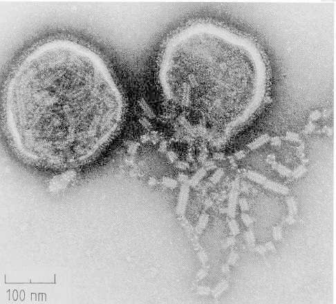
Parainfluenza virus type 1, Sendai strain
2026-02-21 14:23:36
Fever — usually low-grade to moderate
Rhinorrhoea — watery discharge due to nasal mucosal inflammation
Nasal congestion and blockage
Sore throat (pharyngitis)
Dry cough — early stage
Hoarseness of voice — laryngeal involvement
Barking cough — characteristic of croup (laryngotracheobronchitis)
Noisy breathing (stridor) — especially in children
Breathing difficulty — due to upper airway inflammation
General malaise and fatigue
Headache and body ache (mild)
Reduced appetite in children
Infants and young children may present primarily with:
Irritability
Feeding difficulty
Sleep disturbance due to cough and airway obstruction
Supportive management is the mainstay of therapy
No specific antiviral approved for routine use
Rest and adequate oral hydration
Paracetamol for fever and discomfort
Humidified air reduces airway irritation and helps mucus clearance
Nasal saline drops relieve congestion
For croup (laryngotracheobronchitis):
Single-dose oral/IM dexamethasone reduces airway edema
Nebulized adrenaline (epinephrine) for moderate–severe stridor
Oxygen therapy if hypoxic
For bronchiolitis or pneumonia:
Oxygen support as needed
Close monitoring of respiratory effort
IV fluids if poor oral intake
Ventilatory support in severe respiratory failure
Antibiotics only if secondary bacterial infection suspected
Hospital admission for severe airway obstruction, persistent stridor at rest, or respiratory distress
Tap a card to view full section
Use the coloured cards above (Etiology, Symptoms, Treatment, etc.).