Summary / Overview
- RSV (Respiratory Syncytial Virus) is a common contagious virus that causes lower-respiratory tract infections, especially in infants, young children, and older adults.
- It is the leading cause of bronchiolitis and pneumonia in infants worldwide.
- Most infections are mild, but severe disease occurs in premature infants, those with congenital heart/lung disease, immunocompromised adults, and the elderly.
- Key features: seasonal outbreaks, high reinfection rates, and significant hospitalization burden in children <2 years.
Etiology
- RSV is an enveloped, negative-sense, single-stranded RNA virus from the Paramyxoviridae family.
- Belongs to the genus Orthopneumovirus.
Pathogenesis
- RSV primarily targets the epithelial cells lining the small airways (bronchioles).
- Severe RSV infection can cause “syncytia” formation — multinucleated giant cells created by fusion of infected cells.
Symptoms
- Initial symptoms are mild, resembling common cold.
Signs
- Tachypnea and wheezing are hallmark findings.
Clinical Features
- RSV bronchiolitis is most severe in infants <6 months and premature babies.
- Wheezing is the hallmark of RSV lower-respiratory involvement.
- In high-risk infants, RSV can lead to severe hypoxemia requiring hospitalization.
Investigations
- RSV is primarily a clinical diagnosis in otherwise healthy children.
- Chest X-ray is not routinely required; used only in severe or atypical cases.
- RT-PCR is the most sensitive method for RSV detection.
Differential Diagnosis
- RSV must be differentiated from other viral and bacterial causes of lower respiratory tract infection in infants and young children.
- Bacterial pneumonia (Strep pneumoniae, Staph aureus, H. influenzae) – high fever, toxic appearance, lobar consolidation on CXR.
- Congestive heart failure in infants — tachypnea but usually with hepatomegaly and poor feeding.
Complications
- Apnea in young infants — especially <2 months or premature babies.
- Acute respiratory failure — due to bronchiolitis or pneumonia.
- Recurrent wheeze/reactive airway disease — common post-RSV, especially in atopic children.
Treatment
- Supportive care is the mainstay — no specific antiviral for routine RSV cases.
- Mechanical ventilation may be required in severe bronchiolitis or apnea.
- Palivizumab prophylaxis (monthly IM injections) — for high-risk infants only
Prevention
- Strict hand hygiene is the most effective preventive measure.
- Palivizumab prophylaxis for high-risk infants (monthly IM during RSV season)
- Nirsevimab (long-acting monoclonal antibody)
Serotypes / Subtypes
- RSV has two major antigenic subtypes: RSV-A and RSV-B.
- Immunity is incomplete and reinfections are common throughout life.
Pathology
- RSV predominantly infects the epithelial cells of the nasopharynx and lower respiratory tract.
- The hallmark pathological feature is bronchiolitis with edema and narrowing of small airways.
- Severe disease is due to both viral cytopathic effect and an exaggerated host inflammatory response.
Radiology / Imaging
- Chest X-ray in RSV typically shows bilateral hyperinflation and increased peribronchial markings.
- Classic finding: hyperinflated lungs with streaky perihilar opacities.
Notes / Teaching points
- Why does RSV cause bronchiolitis mainly in infants?
- Why is hyperinflation common in RSV?
- Why do some RSV cases develop apnea?
- Why is feeding difficulty a key clinical sign?
- Why not routinely use bronchodilators in RSV?
- Why is bacterial pneumonia uncommon in RSV?
- Why is Palivizumab given only to high-risk infants?
- What is the mechanism of wheeze in RSV?
- Why does RSV season cause epidemics every year?
RSV bronchiolitis is most severe in infants <6 months and premature babies.
• Starts as an upper-respiratory illness (fever, cold, cough) → progresses to lower-respiratory symptoms in 2–4 days.
• Progressive cough becoming harsh or paroxysmal.
• Increasing tachypnea (RR > 60/min in infants).
• Feeding difficulty due to breathlessness.
• Signs of respiratory distress: chest indrawing, nasal flaring, grunting.
Wheezing is the hallmark of RSV lower-respiratory involvement.
• Diffuse crackles (crepitations) on auscultation.
• Air trapping may cause hyperinflated chest.
• Apnea episodes may be the presenting feature in very young infants.
• Cyanosis in severe disease.
In high-risk infants, RSV can lead to severe hypoxemia requiring hospitalization.
Apnea in young infants — especially <2 months or premature babies.
Acute respiratory failure — due to bronchiolitis or pneumonia.
• Severe bronchiolitis → air trapping, atelectasis, respiratory distress
• Dehydration due to poor feeding
• Otitis media (common in infants)
• Secondary bacterial pneumonia (Strep pneumoniae, Staph aureus)
• Acute otitis media
• Wheezing episodes lasting weeks after acute illness
Recurrent wheeze/reactive airway disease — common post-RSV, especially in atopic children.
• Rare: myocarditis, hepatitis
• Rare neurologic complications: seizures, encephalitis
RSV must be differentiated from other viral and bacterial causes of lower respiratory tract infection in infants and young children.
• Influenza A/B – tends to have higher fever, systemic myalgias, abrupt onset.
• Parainfluenza virus – often associated with croup (barking cough, stridor).
• Adenovirus – may cause conjunctivitis, diarrhea, more prolonged fever.
• Human metapneumovirus – clinically similar to RSV; younger infants often more affected.
• Rhinovirus – usually milder, upper respiratory predominance.
• COVID-19 – variable presentation; may show anosmia, older age involvement.
Bacterial pneumonia (Strep pneumoniae, Staph aureus, H. influenzae) – high fever, toxic appearance, lobar consolidation on CXR.
• Chlamydia trachomatis pneumonia in infants – staccato cough, conjunctivitis, eosinophilia.
• Pertussis – paroxysmal cough, inspiratory whoop, lymphocytosis.
Congestive heart failure in infants — tachypnea but usually with hepatomegaly and poor feeding.
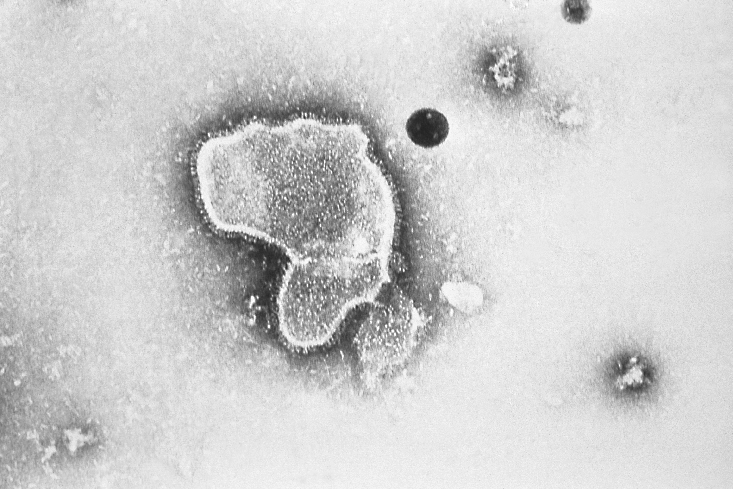
RSV is an enveloped, negative-sense, single-stranded RNA virus from the Paramyxoviridae family.
Belongs to the genus Orthopneumovirus.
Two major antigenic subtypes:
- RSV-A
- RSV-B
RSV-A usually dominates during outbreaks and is more virulent.
RSV infects ciliated epithelial cells of the upper and lower respiratory tract.
Fusion (F) protein causes **syncytia formation**, allowing cell-to-cell spread.
G-protein mediates viral attachment to host cells.
Transmission occurs via respiratory droplets, close contact, and contaminated surfaces.
Virus can survive several hours on surfaces (fomites).
Incubation period: 3–7 days.
RSV is primarily a clinical diagnosis in otherwise healthy children.
• Routine investigations are **NOT needed** for mild typical cases.
• Pulse oximetry — essential to assess oxygen saturation.
Chest X-ray is not routinely required; used only in severe or atypical cases.
• Typical CXR findings: hyperinflation, peribronchial thickening, patchy atelectasis.
• CBC: usually normal or mildly elevated WBC; neutrophilia suggests bacterial coinfection.
• Nasopharyngeal swab for RSV antigen or RT-PCR — mainly used in infants at high risk, hospitalized cases, outbreaks.
RT-PCR is the most sensitive method for RSV detection.
• ABG in severe respiratory distress or persistent hypoxemia.
• Electrolytes if dehydration suspected due to poor feeding.
Why does RSV cause bronchiolitis mainly in infants?
RSV preferentially infects small bronchioles. Infants have narrow airways → minimal edema or mucus causes critical obstruction → respiratory distress.
Why is hyperinflation common in RSV?
Air trapping occurs because inflamed bronchioles collapse during expiration, preventing complete exhalation.
Why do some RSV cases develop apnea?
Immature respiratory control in infants + airway inflammation → transient pauses in breathing. More common in premature infants.
Why is feeding difficulty a key clinical sign?
Nasal obstruction + increased work of breathing → infants cannot coordinate suck–swallow–breathing → poor intake & dehydration.
Why not routinely use bronchodilators in RSV?
RSV pathology is airway inflammation & mucus plugging (not smooth-muscle spasm). Most infants show little or no response.
Why is bacterial pneumonia uncommon in RSV?
RSV primarily involves the small airways (bronchioles) rather than alveoli. If consolidation develops, think secondary bacterial infection.
Why is Palivizumab given only to high-risk infants?
It is expensive monoclonal antibody prophylaxis. Reserved for infants with prematurity, hemodynamic heart disease, or chronic lung disease of prematurity.
What is the mechanism of wheeze in RSV?
Small airway narrowing + mucus → turbulent airflow → expiratory wheeze. Not classical asthma, though later wheezing episodes may increase.
Why does RSV season cause epidemics every year?
RSV is highly contagious, survives on surfaces, spreads by droplets; immunity is incomplete and reinfection is common.
RSV primarily targets the epithelial cells lining the small airways (bronchioles).
• Viral fusion protein (F-protein) allows the virus to enter host respiratory epithelial cells.
• Infection triggers apoptosis, cell sloughing, and necrosis in bronchiolar walls.
• Dead cells + mucus + inflammatory exudate → **bronchiolar plugging**, producing air trapping.
• Narrow bronchioles in infants → **atelectasis**, wheezing, and hypoxia.
• Peribronchiolar infiltration with lymphocytes, neutrophils, and plasma cells.
Severe RSV infection can cause “syncytia” formation — multinucleated giant cells created by fusion of infected cells.
• Inflammation increases airway resistance → respiratory distress.
RSV predominantly infects the epithelial cells of the nasopharynx and lower respiratory tract.
• Causes necrosis and sloughing of bronchiolar epithelium
• Leads to formation of **mucus plugs** composed of debris, fibrin, and inflammatory cells
• Results in **air trapping**, **hyperinflation**, and **atelectasis**
The hallmark pathological feature is bronchiolitis with edema and narrowing of small airways.
• Peribronchiolar lymphocytic infiltration
• Submucosal edema → obstruction
• Giant cell formation may be seen in severe disease
• In infants: small airway size magnifies the obstruction
• In adults/elderly: pathology resembles viral pneumonitis
Severe disease is due to both viral cytopathic effect and an exaggerated host inflammatory response.
Strict hand hygiene is the most effective preventive measure.
• Avoid close contact with coughing/sneezing individuals
• Avoid kissing infants on face during respiratory illness
• Regular cleaning of surfaces, toys, and frequently touched objects
• Avoid cigarette smoke exposure (major risk factor for severe bronchiolitis)
• Encourage breastfeeding — provides maternal antibodies and lowers severity
Palivizumab prophylaxis for high-risk infants (monthly IM during RSV season)
• Preterm < 29 weeks gestation
• Chronic lung disease of prematurity
• Hemodynamically significant congenital heart disease
• Certain immunocompromised infants
Nirsevimab (long-acting monoclonal antibody)
• One-time dose providing season-long protection in infants
• Recommended in many countries for infants entering their first RSV season
• No licensed RSV vaccine for infants yet (as of current guidelines)
• Maternal RSV vaccination under evaluation to protect newborns via passive antibodies
Chest X-ray in RSV typically shows bilateral hyperinflation and increased peribronchial markings.
• Peribronchial cuffing (“donut sign”)
• Patchy atelectasis due to mucus plugging
• Flattened diaphragms from air trapping
• Interstitial infiltrates more common than alveolar consolidation
Classic finding: hyperinflated lungs with streaky perihilar opacities.
• Lobar pneumonia is uncommon (if present, suspect bacterial coinfection)
• In severe bronchiolitis: diffuse haziness with areas of segmental collapse
• CT is rarely required; when done shows small airway inflammation and air-trapping mosaic pattern
RSV has two major antigenic subtypes: RSV-A and RSV-B.
• Both A and B circulate simultaneously each season
• RSV-A usually causes larger outbreaks and is often more severe
• Subtypes are classified based on differences in the G-glycoprotein
• Multiple genotypes exist within each subtype (e.g., ON1 for RSV-A, BA for RSV-B)
Immunity is incomplete and reinfections are common throughout life.
Tachypnea and wheezing are hallmark findings.
• Tachypnea
• Chest indrawing / retractions
• Nasal flaring
• Expiratory wheeze
• Crackles on auscultation
• Prolonged expiratory phase
• Hypoxia (in moderate–severe cases)
*Apnea episodes* — especially in premature infants.
RSV (Respiratory Syncytial Virus) is a common contagious virus that causes lower-respiratory tract infections, especially in infants, young children, and older adults.
It is the leading cause of bronchiolitis and pneumonia in infants worldwide.
Most infections are mild, but severe disease occurs in premature infants, those with congenital heart/lung disease, immunocompromised adults, and the elderly.
Key features: seasonal outbreaks, high reinfection rates, and significant hospitalization burden in children <2 years.
References
CDC (https://phil.cdc.gov/details.aspx?pid=2175)
2025-11-24 13:13:56
Initial symptoms are mild, resembling common cold.
• Low-grade fever
• Nasal congestion / rhinorrhea
• Cough
• Poor feeding (especially infants)
• Irritability
Supportive care is the mainstay — no specific antiviral for routine RSV cases.
• Adequate hydration (oral/NG/IV as needed)
• Frequent nasal suctioning (bulb suction or saline instillation)
• Antipyretics (paracetamol/ibuprofen — age-appropriate)
• Oxygen therapy if SpO₂ < 90–92%
• Humidified oxygen / HFNC in moderately severe bronchiolitis
Mechanical ventilation may be required in severe bronchiolitis or apnea.
• Nebulized hypertonic saline sometimes used (benefit modest; not routine)
• Bronchodilators (trial in older infants with wheeze; not routinely recommended)
• Corticosteroids — **not routinely recommended** in infants with bronchiolitis
• Antibiotics only if there is **proven** secondary bacterial infection
Palivizumab prophylaxis (monthly IM injections) — for high-risk infants only
• Preterm infants < 29 weeks
• Chronic lung disease of prematurity
• Significant congenital heart disease
Tap a card to view full section
Use the coloured cards above (Etiology, Symptoms, Treatment, etc.).